


Breast fibroids are a common degenerative disease. There are specific symptoms of thoracic osteonecrosis, indicating the onset of pathology. In the early stages, the discomfort does not bother the patient much, so do not rush to seek the help of a specialist. Over time, the symptoms increased, forcing the patient to go to the doctor and discover the neglected pathology. You should look out for the early signs to determine osteonecrosis and what treatment is most effective.
What is thoracic fibroma and how does it arise?
Osteoma of the thoracic region is characterized by the appearance of destructive-dystrophic processes in the middle part of the vertebrae. The destruction is located between the 8th and 19th vertebrae. To find out which vertebra is affected, it is necessary to conduct accurate diagnostic studies. Thoracic osteomas are often associated with formidable complications, including prolapse or hernia. Without complications, the disease is rare, as destruction of cartilage tissue inevitably leads to destruction of the entire vertebral framework.
When a patient has a circulatory disorder or due to age, the joint wears out, the fibrous capsule in the space of the disc begins to collapse, losing its normal structure. Because the destruction takes place slowly, in the early stages, small cracks appear, through which the pulp nucleus penetrates.
As the internal components seep out, the ring fibers begin to weaken, leading to gradual dilation and rupture. When the nucleus pulposus protrudes, disc herniation occurs, which is the most common complication of osteonecrosis. Pathology involves damage to cartilage tissue, causing significant discomfort. Severe back pain is also associated with neurological syndromes that develop as a result of nerve root compression or irritation.
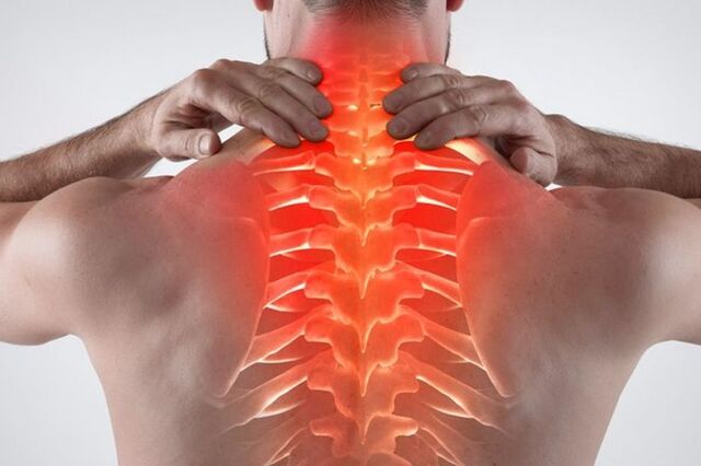
Symptoms of osteonecrosis of the breast
In the early stages, the patient does not feel discomfort, so at this stage, the disease can only be detected by chance. The disease has many symptoms that can be disguised as other diseases.
Symptoms of osteonecrosis of the chest can be felt by the following manifestations:
- Shortness of breath. Problems arise, manifesting as shortness of breath and a feeling of being out of breath. This shows damage to the thoracic vertebrae and spinal cord.
- The main symptom is pain in the chest area. There is also a pressing feeling in the heart, like an ischemic attack.
- Discomfort occurs when the back is bent. As the disease progresses, the pain in this area increases.
- Against the background of impaired blood circulation, there is a feeling of cold in the lower or upper extremities.
- Pain in the chest against the background of an emerging disc herniation. Discomfort is usually felt stronger on the left or right side of the affected area.
- Sore throat and difficulty swallowing. If there is irritation of the nerve endings in the upper part of the thoracic region, a cough will occur.
- Women may experience breast pain that is not related to cyclical changes or hormonal imbalances.
- Tingling or burning sensation in the legs and feet.
- Hair and nails become brittle and dull.
- Herpes zoster is less common.
- Back and chest pain occur at the same time.
- Less commonly, there is discomfort in the stomach, liver or pancreas.
- Onset of stiff pain in the ribs, manifestation of intercostal neuralgia.
- There are signs of fibrosis and thoracic compression - a similar pathology.
- There are problems in the work of the digestive tract. Feeling nauseous, heavy stomach.
- In men, some libido may drop. Problems arise in the genital area.
- When standing or sitting for a long time, severe discomfort occurs.
- Severe headache accompanied by dizziness. Migraine headaches with aura may be present.
- Patients often present with intercostal neuralgia.
- The pain may radiate to the neck or lower back.
If you find an osteosarcoma in the thorax and its signs or some of its signs, you should urgently consult a therapist, neurologist, orthopedist. In addition, such symptoms should be warned in the absence of problems with the gastrointestinal tract, cardiovascular system and lungs.
There are also acute and subacute symptoms. If, with an exacerbation of osteonecrosis of the thoracic region, the patient has severe pain that makes the patient unable to work and can only rest in bed, the subacute development is slow and does not significantly limit activitiespatient movement.
A clear sign of a sluggish lesion - no acute pain. Symptoms in the subacute phase are cleared. No discomfort with basic body movements, including inhaling, sneezing, or turning. A person does not experience pain in a dream, so the process of falling asleep is facilitated.
In order for the subacute course of the disease not to worsen and go into remission, it is necessary to observe important rules:
- Prohibit lifting weights.
- You cannot bend over quickly.
- Do not stay in a sitting or standing position for a long time. A person often unconsciously in this state assumes a posture that is harmful to the spine, so there is an excessive load on the ribs, which leads to another exacerbation.
- Avoid hypothermia. It has been proven that failure to adhere to a comfortable temperature regime for the body becomes an exacerbation of the inflammatory process. Moisture is also bad for joints.
The duration of the subacute course is individual. If medical recommendations are followed, the patient will completely get rid of discomfort within 2-3 weeks. If conservative treatment and rest do not help, and the patient begins to experience nausea, dizziness, and weakness, urgent medical attention is needed. Such symptoms indicate a recurrent exacerbation.
Degree of development of thoracic osteonecrosis
There are 4 clinical stages of the disease, during which the patient develops signs of pathology:
- In the early stages, there are no clinical symptoms. The first stage occurs against the background of the appearance of destructive processes in cartilage and bone tissue. In the early stages, the annulus is not broken or stretched, so there is no hernia. They can detect the initial protrusion and signs of cartilage degeneration.
- The second stage presents with mild pain or discomfort. The patient looked for a doctor attentively, so the thoracic osteonecrosis was detected in time. Patients who do not want to visit a specialist can still suffer in stage 2, using available remedies but buying medicine for a long time will not be enough. At this stage, the most common neurological symptoms may appear, including headache, interstitial burning, neck pain, and spikes in blood pressure. Also at this stage, there is an increase in degenerative damage in the spine: the fibrous ring protrudes out leading to the appearance of a herniated disc in the thoracic region.
- The third stage is inherently difficult for the patient. Persistent neurologic syndromes develop, including persistent radiating pain in the shoulder blades, arms, collarbone, and lower back. Patients may present with somatic and autonomic disturbances, indicating disturbances in nervous system functioning. Patients often suffer from nausea, headaches, dizziness, back pain. Cardiac, digestive, or pulmonary disfiguring signs of the disease may also be present. At this stage, there is an active demineralization of bone and cartilage tissue. Tendency to trauma.
- The final stage is the fourth stage. Against the background of osteochondrosis and hernia, irreversible consequences arise - the mobility of the intervertebral discs is completely lost, and the cartilage tissue in the place of long-standing inflammation is replaced by osteoblasts. To remove them, an operation is required.
To keep your body from going into a state similar to stage 3 or 4, it's better to see your doctor at the slightest sign. The sooner the disease is detected and treated, the quicker the patient will return to normal and learn to live with osteonecrosis. The process of pathological destruction cannot be completely stopped, but it can be slowed down by leading a healthy lifestyle, using medications and performing daily exercises. The later a patient arrives to see a doctor, the harder it is to get rid of the severe pain syndrome associated with cartilage degeneration.
Risk factors and causes of the disease
There is no exact reason that causes destructive changes in the spine. An important role in the occurrence of pathology is thought to be due to genetic factors. It has been proven that people who are not physically active are more likely to have spine problems than people who exercise regularly. In addition, excessive physical activity can cause cartilage destruction at an early age.
The thinning and destruction of discs is closely related to spinal overload. If the muscles are not strong enough, and the back is subjected to frequent overload, it will lead to the destruction of cartilage tissue.
What are the possible causes of osteonecrosis:
- Fat. When you are overweight, there is a lot of weight pressure on the spine. As a result, premature bone tissue destruction occurs.
- The presence of abnormalities in the structure of bones and cartilage. Such problems arise even during the period of intrauterine development.
- Congenital asymmetry of interarticular spaces in intervertebral disc joints belongs to the type of tropical anomalies, which contributes to the occurrence of degenerative-dystrophic processes in the spine.
- The presence of muscle spasms, spondylolisthesis, chronic persistent trigger points and vascular disorders in the thoracic region. These pathologies also contribute to the occurrence of osteonecrosis of the thoracic region.
- Prolonged exposure to vibrations on the spine in a sitting position. An example of a job is a minibus or bus driver.
- Physical stress is often associated with heavy lifting. Examples include work as a porter or professional sports activities.
- Smoking and alcohol abuse. People with an unhealthy lifestyle are prone to mineral deficiencies in the body, poor blood circulation leading to back diseases.
- Sedentary lifestyle. With insufficient physical activity, calcium is washed away more quickly, which is associated with poor metabolism. As a result, the bones become brittle. In addition, muscle tissue atrophies, as a result of which the load on the spine is greatly increased. The result is pain, frequent discomfort with minimal exertion.
Because the intervertebral discs have enough mobility of the intervertebral discs. The discs act as shock absorbers. With the development of osteonecrosis, accelerated demineralization occurs, vital moisture from the joints is lost. This leads to discomfort, reduced mobility of the spine.
Risk factors for osteonecrosis of the breast include:
- Elderly age. In the elderly, a natural degenerative phenomenon occurs, so after the age of 40, the disease is detected more often.
- Female. In girls, there are periods that contribute to the active leaching of calcium from the bones - pregnancy and menopause. If there is no adequate support medicine, it is easy to get diseases of the spine.
- The presence of hormonal disorders, endocrine diseases. If the patient has diabetes or uncompensated hypothyroidism, disc degeneration can occur at an early age.
- Prolonged immobility. If the patient is sick and has to lie down for a long time, the muscles will atrophy, thereby causing back pain.
- Previous back injury. When the ligaments and tendons are stretched, the risk of osteonecrosis in the chest area increases.
- Presence of scoliosis. Poor posture in the future can cause serious spinal problems, including osteonecrosis and hernia.
Diagnosis of thoracic bone tumor
If the patient suspects back problems, a specialist should be consulted. The doctor examines the patient, asks questions, and measures blood pressure. If a neurological problem is suspected, the patient is referred to a narrow specialist - a traumatologist, neurologist or orthopedist.
At the specialist's appointment, they also inquire about the complaint, making an initial diagnosis for the patient. Based on visual examination, a variety of diagnostic measures are prescribed, including:
- X-ray. With the help of X-rays, you can evaluate the condition of the skeletal system in general. If the patient has a herniated or necrotic bone, signs of pathology may be noticed - the distance between the discs will decrease and sometimes a darkening of the color will be noticed at the site of the presumed hernia. If the results of the imaging do not suit the specialist, you must continue to look for the cause of the pain and discomfort.
- CT or MRI. The most accurate diagnostic methods allow you to accurately check the state of the inflammatory foci in the image. More detailed images can be seen on MRI, but if there are contraindications (presence of a pacemaker or prosthetic in the joint), computed tomography is indicated. CT is an innovative X-ray method that allows you to view bones, tendons, and ligaments in detail. The image renders the image as a three-dimensional image, so the details of the damage are clearly visible.
- General blood and biochemical tests. These analyzes are necessary to assess the patient's health. If an increase in leukocytes, ESR is found, then this indicates an active inflammatory process in the body. With active destruction of bone tissue, the amount of calcium decreases, and a deficiency of cholecalciferol (vitamin D3) is found in the blood.
- Spine scan. Research methods show active destruction of bone tissue. Weak bone tissue is easily broken. The method will reveal trends and signs of deterioration.
To diagnose the disease, you need to see an experienced specialist. For a final diagnosis, a complete clinical picture is required, taking into account several laboratory research methods.
Fibroids of the thoracic spine need to be differentiated with the following pathologies:
- Cervical spondylosis.
- Diseases of the urinary system, including urolithiasis, cystitis or pyelonephritis.
- Diseases of the cardiovascular system, except sinus arrhythmias, tachycardia and angina attacks.
- Gastrointestinal diseases, including chronic pancreatitis, gastric and duodenal ulcers, irritable bowel syndrome.
- Previous trauma or fracture.
- Tumors in the chest, including a malignant process.
- Rheumatoid arthritis (determined by blood tests for C-reactive protein, rheumatoid tests, and ESR).
- Inflammation of the spinal cord.
- Acute inflammatory process.
- Ankylosing spondylitis.
- Degenerative spine.
Treatment of osteonecrosis of the thoracic spine
To slow the progression of the disease, an integrated therapeutic approach is required. At the initial stage, only conservative treatment is carried out, including the use of drugs and physiotherapeutic methods for treatment. In severe cases, when the patient has a large hernia and a marked degree of bone degeneration, surgery is indicated. Do not self-medicate at home. Folk remedies do not eliminate osteonecrosis of the thoracic spine.
In what cases is surgery performed?
Osteonecrosis of the thoracic region has a negative impact on the patient's quality of life. If the patient has persistent discomfort that interferes with normal life, due to the ineffectiveness of medication, surgical solutions to the problem may be offered.
Absolute indications for surgery include:
- Lack of sensitivity in the bladder and intestines.
- If the sensitivity in the legs disappears and the patient loses the ability to move independently.
- Paralysis due to overgrowth of hernia.
In other cases, the patient makes the decision to remove the skull mass independently. If the disease is really causing severe pain and the patient's condition has not improved against the background of conservative treatment, doctors recommend surgery.
Drugs to treat osteonecrosis of the thoracic spine
During the exacerbation period, the attending physician prescribes various drugs that are necessary to use to reduce the inflammatory process. The acute phase is characterized by severe pain that can only be relieved by medication. If enough medicine is taken, the patient will get better. Only an experienced specialist can prescribe the drug; self-medication is unacceptable.
Tumors of the thoracic spine are treated with the following drugs:
- Non-steroidal anti-inflammatory drugs, pain relievers, or pain relievers. These medications are designed to provide quick relief from back pain associated with an active inflammatory process. The effect of taking pills or injections will be felt the next day. Taking any drug of the NSAID class is accompanied by side effects with long-term use, so experts recommend limiting its use to a minimum period, no more than 1-2 weeks. Analgesics are most detrimental to the gastric mucosa, causing gastric pathology and inflammation. Patients at risk are given certain drugs designed to protect the gastrointestinal mucosa. Examples include proton pump inhibitors, histamine H2 receptor blockers, antacids. People with ulcers and gastritis are better off avoiding the use of NSAIDs or taking modern analogues with selective action.
- Muscle relaxants. These drugs are very effective in treating spasticity. Relieve pain from muscle tension. They act on trigger points located in the compressed muscle tissue. The more a person tries too hard, the higher their number is. Muscle relaxants provide good relief from muscle tension and thus have a pain-relieving effect. You need to use the drug in a course, the average treatment time is at least 2-4 weeks.
- Group B vitamins. Indications B1, B6, B12 in the form of injections with combination preparations. In large doses, these substances have an analgesic effect and have a positive effect on the nervous system. Neuromodulators are effective in treating pain associated with pinched nerve roots. With the help of nutrition, it is not possible to replenish the norm of these substances necessary to achieve a therapeutic effect, therefore they are prescribed in the form of drugs. The average duration of an injection is 2-3 weeks. Then, if necessary, they switch to tablets.
- Ointments, anti-inflammatory gels. If the pain is tolerable and systemic forms of NSAIDs are contraindicated, then external drugs are prescribed. The advantage of external remedies is that they do not cause side effects. In rare cases, skin allergies may occur, but the ointment will not degrade the gastrointestinal tract or blood in the laboratory. Another advantage of outdoor products is the possibility of long-term use. You can apply the gels for up to 4 weeks, after which they will take a break. The program and duration of treatment are determined by the attending physician.
- Honroprotectors. These are complex substances that are used to nourish the cartilage tissue of the joints. It is necessary to use the drug for a long time, at least six months, then take a break of 2-3 months and repeat the course. Within 2-3 months, injectable release forms are used, as they are better absorbed. They then switched to supportive treatment, including the use of tablets. It is important to understand that the drug does not prevent the destruction of cartilage tissue. They only create additional nutrition, slow down the degenerative process that occurs in bones and joints.
- Complex preparations of calcium and vitamin D3. It has been shown that residents in northern latitudes do not get enough vitamin D3, because year-round solar activity in this region is low. To get rid of anemia, it is necessary to supplement with cholecalciferol in winter and autumn while solar activity is minimal. Without this vitamin, the assimilation of calcium and other minerals is impossible. Due to prolonged calcium deficiency, bone tissue thins over time, as a result of which a person develops osteonecrosis and other complications. Calcium and D3 are better absorbed in combination, therefore complex preparations are prescribed. Dosage and administration should be determined by the attending physician.
To aid in treatment, you can prescribe homeopathic medicines, antispasmodics and complex multivitamins.
Conservative therapy for osteonecrosis of the breast
During the recovery period, the patient should pay attention to full rehabilitation. The more carefully the patient takes care of their health, the less likely the disease will occur.
The most effective conservative treatments include:
- Exercise therapy. With the help of exercises, the patient learns to keep the back straight, strengthens the muscle corset. Physical therapy can be done at any age, several times a week. The complex is selected individually, taking into account the anatomical features of the patient. Start doing it gradually, at first for no more than 5 minutes a day. As physical qualities improve, patients learn to perform more difficult exercises for a longer period of time.
- Support bra. Anatomical devices serve to support weakened muscles, if there are contraindications to strengthening them. The patient chooses the bandage according to the height and type of indication. The attending physician must select the appropriate model. Duration and style of wear are determined individually. You can't wear a corset around the clock, otherwise your back muscles will become even weaker.
- Massage. In medical practice, massage is one of the most common and at the same time effective in conservative treatment, in the case of patients with thoracic osteonecrosis. During recovery, the muscles need extra support. It is helpful when blood flow is temporarily improved and overactive muscles are not branched if the correct technique is used. You need to attend specialist sessions several times a year in courses.
- Physical therapy. Physiotherapy procedures are widespread in trauma, orthopedic, and neurological practices. With the help of procedures, local blood flow is improved, systemic drugs are used externally, and the apparatus acts on damaged tissues. As a result, the muscles are warmed up and the chronic inflammatory process in the affected area is eliminated. Examples of medical procedures - magnetic therapy, shock wave therapy, electrophoresis.
Less commonly, manual therapy and acupuncture are indicated.
Thoracic osteosarcoma is a dangerous disease to begin with. In order to prevent the acute progression of the disease, comprehensive treatment of the pathology is required.